Table Of Content
- Testing the inner ear
- Exfoliation Products
- Sesn2/AMPK/mTOR signaling mediates balance between survival and apoptosis in sensory hair cells under stress
- Why and How to Do a Scalp Detox
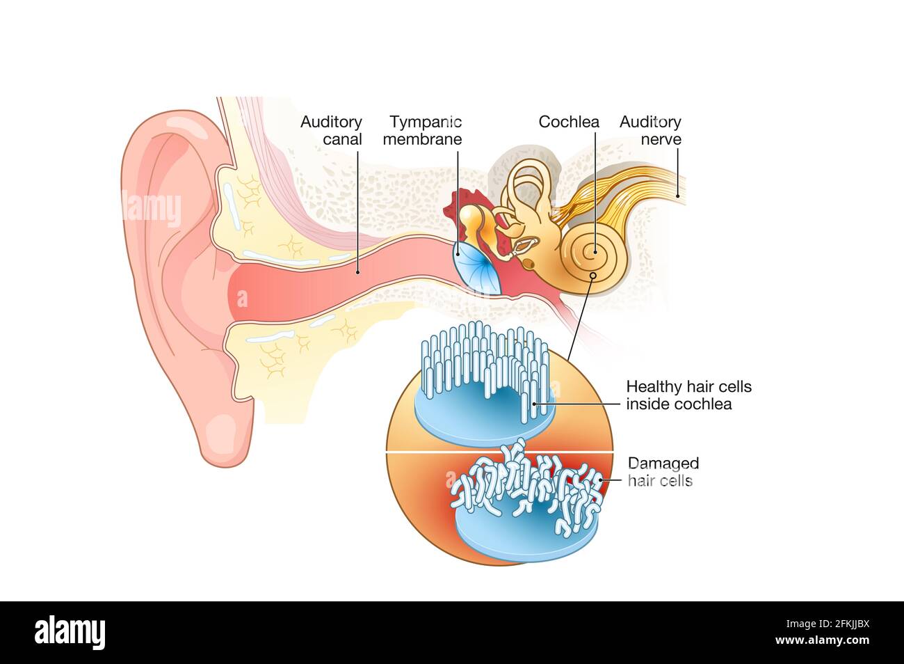
- Reversing hearing loss by regrowing hairs
- Chapter 36: Plant Responses to the Environment
- The 11 Best Hair Growth Serums for Long, Strong Hair
The OHCs are located on the lateral (non-neural) side and are mainly responsible for amplifying acoustic vibrations through periodic contraction and elongation of the cell body driven by changes in membrane potential. The IHCs are located on the medial (neural) side, where they integrate and transmit sound signals to neurons. Synergy between these two types of cells greatly improves the resolution and sensitivity of sound signal processing (Fettiplace, 2017).
Testing the inner ear
The intent of this review is to summarize the more significant structural features and some of the more interesting and important physiological mechanisms that have been elucidated thus far. Outside vertebrates, hair cells are only known to exist in the coronal organ of tunicates. Electrical resonance, electromotility, and their exquisite mechanical sensitivity all contribute to the attractiveness of hair cells as a research subject. Primary cilia are evolutionarily conserved and highly specialized organelles that protrude from cell membranes. Mutations in genes encoding ciliary proteins can cause structural and functional ciliary defects and consequently multiple diseases, collectively termed ciliopathies. The mammalian auditory system is responsible for perceiving external sound stimuli that are ultimately processed in the brain through a series of physical and biochemical reactions.
Exfoliation Products
Neurotransmitter release at the afferent synapse is Ca2+-dependent, as at other neuronal synapses, but there are several important specializations in the hair cell synapse. The release of neurotransmitter is graded in relation to membrane depolarization and the relationship of transmitter release to Ca2+ influx is linear (Glowatzki et al. 2008; Rutherford and Pangrsic 2012; Fettiplace 2017). However it occurs, the net effect of the observed linear relationship at the ribbon synapse is to make transmitter release more sensitive to small depolarizations, thereby enhancing the overall sensitivity of the system. Sensory hair cells are specialized secondary sensory cells that mediate our senses of hearing, balance, linear acceleration, and angular acceleration (head rotation). Sensory hair cells share many structural and functional features across all vertebrate groups, while at the same time they are specialized for employment in a wide variety of sensory tasks. The complexity of hair cell structure is large, and the diversity of hair cell applications in sensory systems exceeds that seen for most, if not all, sensory cell types.
Sesn2/AMPK/mTOR signaling mediates balance between survival and apoptosis in sensory hair cells under stress
Kinocilium develops before stereocilia, finally leading the hair bundle facing toward the non-neural side. Accordingly, the HCs also acquire PCP in readiness for hearing and receiving external stimuli. A, Cross-section of the cochlea (top inset shows diagram of the whole cochlea and sectioning scheme). The three compartments composing the cochlea are indicated (scala timpani, scala vestibuli and scala media).
Why and How to Do a Scalp Detox
These channels belong to the SK2 family of potassium channels, which are sensitive to micromolar concentrations of Ca2+ and insensitive to voltage. Once activated, these Ca2+-activated K+ channels remain open for a long time, due to the time required to bring intracellular Ca2+ down below the concentration that activates them (Fuchs and Murrow 1992; Rohmann et al. 2015). Hair cells of the semicircular canals are located in three ampullae, one for each of the canals. Rotation of the head induces inertial pressure by the fluid within the canal against the cupula, which in turn causes displacement of the hair bundle (Goldberg et al. 2012). As an example of the sensory capability of the semicircular canal system, a housecat is able to right itself and land gracefully after being dropped from an upside-down position in less than the time it takes to fall 1.5 m. Our understanding of the cellular mechanisms of hair cell mechanotransduction has grown rapidly in the past decade.
The researchers showed that Bmp7 promotes the development of low-frequency-sensing hair cells. When they bathed developing basilar papillas in a solution containing Bmp7, they found that all the hair cells—even those at the high-frequency end—developed characteristics of low-frequency-sensing hair cells. These results suggest that during embryonic development, high levels of Bmp7 at one end of the basilar papilla signal the formation of low-frequency-sensing hair cells. Decreasing levels of Bmp7 along the length of the basilar papilla map result in a gradual tuning to higher frequencies. A scalp scrub can help remove excess oils, dead skin cells, and product buildup that pulls up on the scalp and the hair roots. Products are designed for specific hair types, including dry, oily, sensitive, and color-treated.
Unlocking Auditory Resilience: How Our Hearing Cells Self-Repair - Neuroscience News
Unlocking Auditory Resilience: How Our Hearing Cells Self-Repair.
Posted: Mon, 10 Jul 2023 07:00:00 GMT [source]

The second mammalian innovation is the introduction of a second type of hair cell to amplify the basilar membrane’s oscillation at specific sound frequencies. The cochleas of eutherian mammals comprise one row of primary sensory hair cells (inner hair cells, IHCs) and three rows of modulatory hair cells with little or no afferent function (outer hair cells, OHCs). Each inner hair cell receives afferent synapses from 10 to 15 primary afferent nerve fibers, which amounts to 90–95% of the primary afferent fibers.
AdipoRon reduces cisplatin-induced ototoxicity in hair cells:possible relation to the regulation of mitochondrial biogenesis - ScienceDirect.com
AdipoRon reduces cisplatin-induced ototoxicity in hair cells:possible relation to the regulation of mitochondrial biogenesis.
Posted: Wed, 10 Jan 2024 08:00:00 GMT [source]
In the presence of tones that match the resonant frequency range of the cell, this would yield enhanced receptor potentials compared to a cell lacking resonance (Wu et al. 1995). It is now well-established that CDH23 forms the upper region of the tip link and PCDH15 forms the lower (Zhao and Müller 2015). Subsequent searches, based on genes responsible for a severe deafness syndrome (Usher Syndrome Type 1, or USH1), have led to the description of proteins that engage with CDH23 or PCDH15 and could possibly form the mechanotransducer channel. These searches were productive and yielded important components of the stereocilia tip-link complex, including harmonin, sans, and myosin VIIa, which form a complex at the upper end of the tip-link and interact with CDH23. Also identified were CIB2, TMIE, and LHFPL5 (also known as TMHS), which form a complex at the lower end of the tip link and interact with PCDH15 (Zhao and Müller 2015). Both TMIE and LHFPL5 are integral membrane proteins and could possibly contribute to a transmembrane ion channel.
The researchers called this group the epidermal growth factor, or EGF, which switches on support cells in the auditory system of birds. These support cells then spark the production of new sensory hair cells. Each hair cell also has a connection to the hearing nerve, but the inner hair cell is most responsible for sending sound through the hearing nerve to the brain.
Recent progress has been reviewed in depth by Fettiplace and Kim (2014) and Fettiplace (2017). This influx of positive charge depolarizes the cell, increasing the voltage across the membrane. This causes voltage-gated calcium (Ca2+) channels in the cell body to open, and Ca2+ flows into the cell. Ca2+ triggers a signaling cascade that causes synaptic vesicles containing excitatory neurotransmitter molecules to fuse to the cell membrane and be released, exciting the postsynaptic auditory nerve cell and increasing the transmission of action potentials to the brain. When the stereocilia are pushed in the opposite direction, towards the shortest stereocilia, the tip links relax, the cation channels close, and the cell becomes hyperpolarized (i.e., the membrane potential is more negative) compared to its resting state.
The answer seems to be a combination of increased overall sensitivity to sound volume and decreased sensitivity to tone. This was initially demonstrated by stimulating efferent axons while recording the electrical responses to pure tone acoustic stimuli in hair cells of the turtle cochlea (Art et al. 1985). This experimental paradigm allowed identification of the characteristic resonant frequency of the hair cell. At the same time, however, the hair cell response to lower frequencies was enhanced, but in a flat, frequency-independent manner. This can be explained by an overall increase in the cell input resistance, as the hyperpolarization induced by the efferent synapse caused closure of voltage-gated K+ and voltage-gated Ca2+ channels that were open in the resting cell (Fuchs and Parsons 2006). In the extracellular space, adjacent stereocilia are linked together by a variety of connections (Goodyear et al. 2005; Fettiplace and Kim 2014).
Shortening of the OHC is accompanied by proportional increase of cell radius; the cell volume remains constant. Shortening of the OHC under natural conditions begins with displacement of the stereocilia bundle and opening of mechanosensitive channels at their tips. Depolarization-induced shortening results from activation of a piezoelectric effect, meaning that electrical depolarization and physical shortening are directly coupled.


No comments:
Post a Comment